
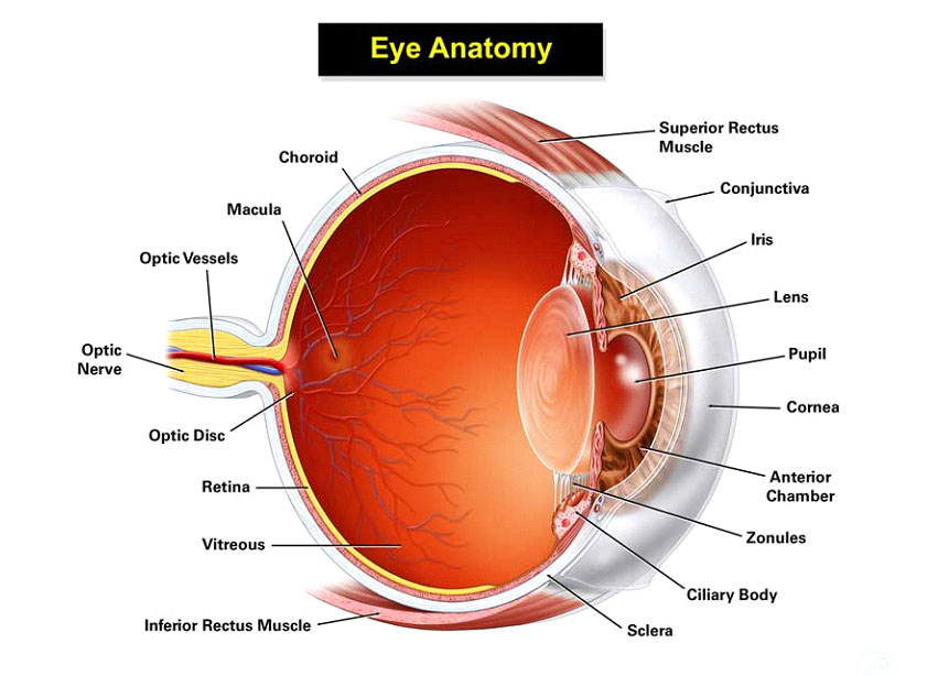
Conjunctiva
The conjunctiva is a transparent mucous membrane that covers the inner surface of the eyelids and the surface of the eye.When it is inflamed or infected it becomes red or pink.This is called conjunctivitis or “pinkeye”.
Sclera
The sclera is the white outer wall of the eye.It covers nearly the entire surface of the eyeball.It is a strong layer made of collagen fibers.The tendons of the six extraocular muscles attach to the sclera.
Cornea
The cornea occupies the front center part of the outer wall of the eye.It is made of collagen fibers in a very special arrangement so that the cornea is clear. One looks through the cornea to see the iris and pupil.The cornea bends light coming into the eye so that it is focused on the retina.The cornea is the part of the eye on which contact lenses are placed.
Iris/Pupil
The iris is the colored part of the eye. It is disc shaped with a hole in the middle (the pupil). Muscles in the iris cause the pupil to constrict in bright light and to dilate in dim light. The change in pupil size regulates the amount of light that reaches the posterior (back) part of the eye.
Lens
The lens of the eye is located directly behind the pupil. The lens bends light coming into the eye to help focus it on the retina. It changes shape to help the eye focus to see objects clearly at near. The lens is suspended from the wall of the eye by many small fibers (zonules) that attach to its capsule.
Ciliary Body
The ciliary body is attached to the outer edge of the iris near the wall of the eye. The ciliary body produces the fluid (aqueous humor) that fills the eye and nourishes its structures. It also helps to change the shape of the lens when focusing occurs.
Viteous
The vitreous cavity lies between the lens and the retina and fills 4/5 of the space inside the back part of the eye.A gelatinous substance known as the vitreous humor fills the cavity.This plays an important role in nourishing the inner structures of the eye.Light comes into the eye through the pupil and passes through the vitreous to be projected on the retina.
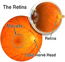
The retina is a thin, transparent structure that covers the inner wall of the eye. The eye works like a camera, and the retina is similar to flim in camera.It is where images are first projected before they are transmitted through the optic nerve to the brain. It is a very complex structure with 10 layers of specialized cells including the photoreceptor cells (rods and cones).
Refractive Errors
Myopiya
The optic nerve connects each eye to the brain.It is a structure that sends the picture seen by the eye to the brain so that it can be processed.The optic nerves end in a structure called the optic chiasm. In an adult, the optic nerve is about the diameter of a pencil.There are over 1 million individual nerve cells in the optic nerve.
Hyperopiya
Farsightedness means it’s easy to see things that are far away, but your close-up vision (near vision) is blurry. The technical term for farsightedness is hyperopia.
Common Eye Diseases
Astigmatism
Astigmatism is a common vision problem caused by an error in the shape of the cornea. With astigmatism, the lens of the eye or the cornea, which is the front surface of the eye, has an irregular curve. This can change the way light passes, or refracts, to your retina. This causes blurry, fuzzy, or distorted vision.
Presbyopiya
 Presbyopia is an eye condition in which your eye slowly loses the ability to quickly focus on objects that are close.It is a disorder that affects everyone people during the natural aging process of their eyes.
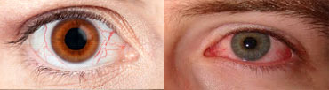
Conjunctivitis
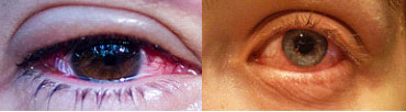 Conjunctivitis, which is commonly called “pink eye”, is an infection or swelling in the eye area. Blood vessels in the conjunctiva, a thin membrane that lines part of the eye, become inflamed.This gives the eye a red or pink color that’s commonly associated with conjunctivitis.
Blepharitis
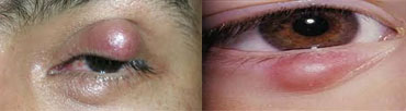 Your eyelids are the folds of skin that cover your eyes and protect them from debris and injury. Your eyelids also have lashes with short, curved hair follicles on the edge of the lids. These follicles contain oil glands. These oil glands can sometimes become clogged or irritated, which triggers inflammation. This condition is known as eyelid inflammation.
Dry Eyes
 Dry eyes occur when your eyes do not produce enough tears, or produce tears that can’t effectively keep your eyes moist. Tears are needed to help keep enough moisture in your eyes.They keep the eye surface smooth, wash away foreign materials, and also help to protect the eyes from infection.
Pterygium
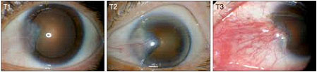 A pterygium is a growth that develops on the conjunctiva or mucous membrane that covers the white part of your eye. It’s a benign or noncancerous growth that’s often shaped like a wedge. In some cases, a pterygium can extend to the cornea. This is the clear part of your eye that covers your iris and pupil. A pterygium usually doesn’t cause problems or require treatment, but it can be removed if it interferes with your vision.
Strabismus
Crossed eyes, also called strabismus, is a condition in which your eyes do not line up properly. If you have this condition, your eyes look in different directions, with each eye focusing on a different object. The disorder is very common in children, affecting four percent of children age 6 and younger. Its cause at birth is not known, but it does tend to run in families. In adults, the disorder can be caused by a variety of factors, including a brain tumor, retina damage, diabetes, or a stroke. Crossed eyes can usually be corrected with eyeglasses and/or surgery.
Diabetic Retinopathy
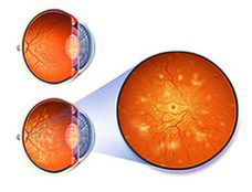 Diabetic retinopathy is a condition that occurs as a result of damaged blood vessels of the retina in people who have diabetes. Diabetic retinopathy can develop whether you have type 1 or type 2 diabetes. While you may start out with only mild vision problems, you can eventually go blind. It is also the most common disease of the eye in diabetics.
Glaucoma
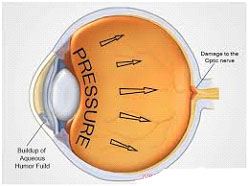 Glaucoma refers to a group of related eye disorders that all cause damage to the optic nerve that carries information from the eye to the brain. Glaucoma usually has few or no initial symptoms.In most cases, glaucoma is associated with higher-than-normal pressure inside the eye – a condition called ocular hypertension. But it also can occur when intraocular pressure (IOP) is normal. If untreated or uncontrolled, glaucoma first causes peripheral vision loss and eventually can lead to blindness.